
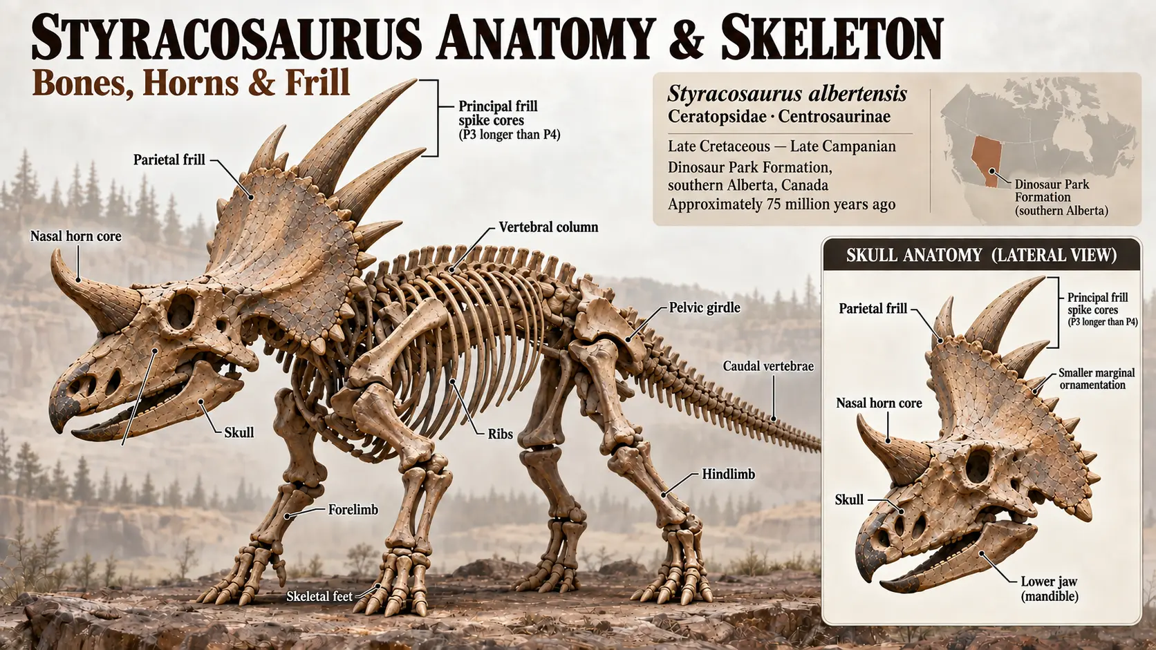
Styracosaurus was a centrosaurine ceratopsid with a robust quadrupedal skeleton, a large skull, an elongate nasal horn core, and a broad parietosquamosal frill bearing prominent marginal processes. Most detailed anatomical knowledge of Styracosaurus albertensis comes from the holotype CMN 344 and additional skull and postcranial specimens from the upper Dinosaur Park Formation of Alberta.
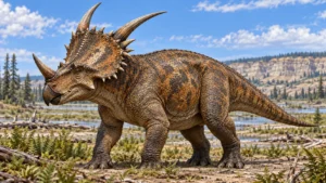
The holotype preserves a largely complete skull together with most of the postcranial skeleton, making CMN 344 the principal anatomical reference for the species. Later studies of additional specimens have expanded knowledge of skull variation, ontogeny, and asymmetry.
At a Glance
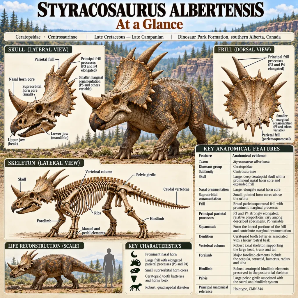
| Feature | Anatomical evidence |
|---|---|
| Taxon | Styracosaurus albertensis |
| Dinosaur group | Ceratopsidae |
| Subfamily | Centrosaurinae |
| Skull | Large, deep ceratopsid skull with a prominent nasal horncore and expanded frill |
| Nasal ornamentation | Large, elongate nasal horncore |
| Supraorbital ornamentation | Small, pointed horncores above the orbits |
| Frill | Broad parietosquamosal frill with prominent marginal processes |
| Principal parietal processes | P3 and P4 strongly elongated in characteristic specimens; P5 variable |
| Squamosals | Form the lateral portions of the frill and contribute marginal ornamentation |
| Dentition | Ceratopsid tooth batteries associated with a horny rostral beak |
| Vertebral column | Robust axial skeleton supporting the large head, trunk and tail |
| Forelimb | Scapula, coracoid, humerus, radius and ulna preserved in the holotype |
| Hindlimb | Robust ceratopsid hindlimb elements preserved in the postcranial skeleton |
| Pelvis | Large pelvic girdle associated with the sacral and hindlimb system |
| Principal anatomical reference | Holotype CMN 344 |
Skull Anatomy
The skull is the most distinctive part of the Styracosaurus skeleton. CMN 344 preserves much of the skull, although some areas were damaged or reconstructed during preparation.
The skull features an elongated nasal region, a large nasal horncore, small supraorbital horncores, and an expanded posterior frill. The skull roof contains the large temporal openings characteristic of ceratopsids, while the cheek region incorporates the jugal, quadratojugal, and squamosal bones.
The original description of CMN 344 emphasized the unusual nasal horn and the series of large processes along the posterior margin of the frill. Subsequent anatomical work has shown that the apparent completeness of the original mounted skull should not be taken literally: portions of the specimen were reconstructed in plaster, and some elements of the rostrum, lower jaws, and frill were incomplete.
Major Skull Regions
| Region | Anatomical significance |
|---|---|
| Nasals | Form the dorsal portion of the snout and support the large nasal horncore |
| Premaxillae | Form the front of the snout and contribute to the rostral region |
| Maxillae | Form much of the tooth-bearing cheek region |
| Frontals and parietals | Contribute to the skull roof and frill |
| Postorbitals | Form the region immediately behind the orbits and bear the small supraorbital ornamentation |
| Jugals | Contribute to the lateral cheek region |
| Squamosals | Form the lateral portions of the frill and contribute marginal ornamentation |
| Parietals | Form the central and posterior portion of the frill |
| Quadrates | Form part of the jaw articulation |
| Dentaries | Form the principal tooth-bearing lower-jaw elements where preserved |
| Epiossifications | Separate or fused marginal elements associated with frill ornamentation |
Nasal Horncore
The nasal horncore is one of the most recognizable skeletal features of Styracosaurus. In mature S. albertensis specimens, it is an elongate, erect structure arising from the nasal region of the skull.
Its morphology contrasts with the much smaller supraorbital horncores. The combination of a prominent nasal horn and relatively small supraorbital ornamentation is characteristic of S. albertensis and contributes to distinguishing it from other centrosaurines.
The horncore should be treated as a bony structure rather than as evidence for the exact dimensions or appearance of any external keratinous covering.
Supraorbital Ornamentation
Small horncores occur above the orbits. Compared with the large nasal horn, these structures are relatively short and pointed.
This condition is significant because supraorbital ornamentation varies substantially among centrosaurines. In S. albertensis, the available specimens document the characteristic relatively small condition, although individual skulls differ in other aspects of cranial ornamentation.
Frill Anatomy
The frill is formed principally by the paired parietal and squamosal bones. Its posterior and lateral margins carry a series of projections and scalloped processes.
The most prominent structures in S. albertensis are the parietal processes conventionally designated P3 and P4, with P5 showing greater variation among specimens.
Parietal Region
| Parietal region | Condition in S. albertensis |
|---|---|
| P1 region | Present in characteristic specimens, although preservation and interpretation vary |
| P2 region | Smaller marginal process |
| P3 | Elongate, prominent process |
| P4 | Elongate, prominent process |
| P5 | Variable in development among specimens |
| More posterior processes | Generally less prominent in typical specimens |
The exact appearance of the frill was not identical in every individual. Comparative studies document variation in the length and shape of marginal processes, particularly toward the posterior portion of the parietal.
This variation is important when reconstructing a Styracosaurus skull: a single specimen should not automatically be treated as representing an invariant frill configuration for the entire species.
Squamosal Anatomy
The paired squamosals form much of the lateral portion of the frill. In S. albertensis, the squamosals contribute marginal ornamentation and help form the characteristic lateral outline of the frill.
The number and expression of individual marginal processes should nevertheless be interpreted in light of preservation and individual variation.
Temporal Openings and Skull Architecture
Like other ceratopsids, Styracosaurus possessed large openings in the posterior skull region. These include the supratemporal and lateral temporal fenestrae associated with the skull roof and cheek region.
The arrangement of these openings reflects the highly modified cranial architecture of ceratopsids, in which the skull combines a large facial skeleton, expanded frill, jaw apparatus, and extensive areas associated with muscle attachment.
The presence of large openings should not be interpreted as evidence that the skull was simply a lightweight structure. The surrounding cranial bones formed an integrated structural framework around the major openings and articulations.
Beak and Tooth-Bearing Region
The anterior jaws formed a toothless rostral region associated with a horny beak. Behind this region, the maxillae and dentaries carried continuously replacing tooth batteries characteristic of ceratopsids.
The holotype skull does not preserve every component of the jaws intact. Consequently, anatomical reconstructions of the complete rostrum and lower jaws should distinguish preserved material from reconstructed portions.
The detailed mechanics of feeding and tooth function belong to the dedicated Styracosaurus Diet, Feeding & Jaw Function page rather than this anatomical overview.
Vertebral Column
The postcranial skeleton of S. albertensis is unusually well documented for a centrosaurine ceratopsid. The postcranial study of CMN 344 provides detailed anatomical information on the vertebral column, pectoral girdle, limbs and pelvic region.
The vertebral column consisted of cervical, dorsal, sacral and caudal regions. The cervical series supported the large skull, while the dorsal vertebrae formed the principal axial framework of the trunk. The sacral region formed the structural connection between the vertebral column and pelvic girdle, and the caudal series extended into the substantial tail.
Because individual vertebrae can be incomplete or damaged, reconstructions of the complete series should not imply that every vertebral element is preserved in a single articulated condition.
Pectoral Girdle
The pectoral girdle includes the scapula and coracoid, which together form the shoulder region and articulate with the forelimb.
CMN 344 preserves the right scapula and coracoid sufficiently well for detailed anatomical and biomechanical study. The scapula has an elongated blade, while the coracoid contributes substantially to the glenoid region.
The scapulocoracoid also provides an important anatomical reference for interpreting forelimb articulation. Later range-of-motion research has used the preserved shoulder elements of CMN 344 to investigate possible forelimb mobility.
Forelimb Skeleton
The forelimb consists principally of the humerus, radius and ulna, followed distally by the wrist and manus.
The holotype preserves the major proximal forelimb elements, including:
| Element | Anatomical role |
|---|---|
| Scapula | Main blade of the pectoral girdle |
| Coracoid | Contributes to the shoulder joint |
| Humerus | Upper forelimb bone |
| Radius | One of the paired forearm bones |
| Ulna | Second major forearm bone |
| Wrist and manus | Distal forelimb structures |
The humerus possesses a distinct proximal articular head associated with the glenoid cavity. The radius and ulna form the paired forearm, while the distal limb terminates in the manus.
The anatomical configuration is consistent with the robust, weight-bearing forelimb construction of ceratopsids. Detailed questions concerning limb posture and range of motion are treated separately under locomotion rather than inferred solely from bone shape here.
Pelvic Girdle
The pelvic girdle forms the connection between the axial skeleton and the hindlimbs. It includes the ilium, pubis, and ischium, with the acetabular region forming the principal articulation for the hindlimb.
The ceratopsid pelvis is heavily integrated with the sacral region. The enlarged pelvic structure provided the bony framework for attachment of the hindlimb and associated soft tissues.
As with the forelimb, pelvic morphology should be distinguished from biomechanical interpretations of how the animal moved.
Hindlimb Skeleton
The hindlimb consists of the femur, tibia, fibula, and ankle and foot elements.
The femur is the principal proximal hindlimb bone and articulates with the acetabulum. Distally, the tibia and fibula form the lower leg, followed by the ankle and pes.
The postcranial material of CMN 344 provides an important anatomical basis for reconstructing the hindlimb skeleton of S. albertensis. However, isolated or incomplete elements from other specimens should not automatically be assumed to represent identical morphology without appropriate taxonomic and anatomical assessment.
Manus and Pes
The hands and feet formed the distal portions of the limbs. Their anatomy reflects the robust, weight-bearing quadrupedal skeleton characteristic of ceratopsids.
Individual phalangeal elements can be difficult to interpret when isolated, so complete skeletal reconstructions should distinguish directly preserved elements from inferred articulation.
Detailed discussion of foot posture, gait and limb mechanics belongs to the Styracosaurus Locomotion, Gait & Movement spoke.
Ontogenetic and Individual Variation
The Styracosaurus skeleton was not anatomically identical at every stage of growth.
Subadult specimens demonstrate changes in cranial ornamentation and other skeletal features during ontogeny. Studies of skull variation also document asymmetry between left and right sides in some individuals.
This has two important consequences for anatomical reconstruction:
- A juvenile or subadult skull should not automatically be reconstructed as a miniature adult.
- Differences between specimens should not automatically be interpreted as differences between species.
The variation documented within S. albertensis is one reason specimen-level evidence is important when describing its anatomy.
What the Skeleton Shows — and What It Does Not
| Anatomical observation | Evidence status | Interpretation boundary |
|---|---|---|
| Large nasal horncore | Strong direct skeletal evidence | Establishes the bony horncore, not its complete external covering |
| Small supraorbital horncores | Strong direct evidence | Exact soft-tissue appearance remains uncertain |
| Expanded parietosquamosal frill | Strong direct evidence | Frill function is addressed separately |
| Elongate P3 and P4 processes | Strong direct evidence in diagnostic specimens | Individual expression varies |
| Variable P5 development | Direct comparative evidence | Should not be treated as a fixed species-wide configuration |
| Robust vertebral column | Direct postcranial evidence | Does not alone establish detailed biomechanics |
| Preserved scapula, coracoid and forelimb | Strong direct evidence from CMN 344 | Range of motion requires separate biomechanical analysis |
| Pelvic and hindlimb skeleton | Direct postcranial evidence | |
| Tooth-bearing jaws and rostral beak region | Direct cranial evidence from available material | Feeding mechanics require separate analysis |
| Ontogenetic skeletal differences | Direct comparative evidence | Juvenile morphology should not be treated as adult morphology |
| Cranial asymmetry | Documented in specimens | Asymmetry should not automatically be interpreted as pathology or species-level difference |
Evidence Assessment
| Anatomical topic | Evidence strength | Assessment |
|---|---|---|
| S. albertensis cranial construction | High | Supported by well-preserved cranial material |
| Nasal horncore morphology | High | Repeatedly documented in S. albertensis specimens |
| Supraorbital ornamentation | High | Characteristic relatively small structures are documented |
| Parietal frill anatomy | High | Diagnostic parietal morphology is well documented |
| Individual frill variation | High | Multiple specimens demonstrate variation |
| Postcranial anatomy | High | CMN 344 provides extensive postcranial material |
| Forelimb anatomy | High | Major pectoral and forelimb elements are preserved and described |
| Hindlimb anatomy | High | Postcranial skeleton provides substantial evidence |
| Complete soft-tissue reconstruction | Low | Soft tissues are not directly preserved |
| Exact horn keratin dimensions | Low | Bony horncores do not preserve the complete external sheath |
| Detailed muscle reconstruction | Moderate to limited | Some attachment surfaces are informative, but full soft-tissue reconstruction remains inferential |
| Exact joint range in life | Low to moderate | Anatomical range can be investigated biomechanically, but actual in-life movement is not directly preserved |
| Uniform skull morphology across the species | Low | Individual, ontogenetic and asymmetrical variation are documented |
Current Scientific Understanding
The anatomy of Styracosaurus albertensis is unusually well documented for a centrosaurine ceratopsid. The holotype CMN 344 combines a substantially preserved skull with most of the postcranial skeleton and therefore remains the central anatomical reference for the species.
Its skeleton is characterized by a large, highly ornamented ceratopsid skull, an elongate nasal horncore, relatively small supraorbital horncores, and a broad frill with prominent parietal and squamosal processes. The postcranial skeleton shows the robust axial column, pectoral and pelvic girdles, and weight-bearing limbs expected in a large centrosaurine.
Additional specimens demonstrate that the skull was not anatomically static. The length and development of some frill processes vary among individuals, and ontogenetic as well as asymmetrical variation complicates attempts to produce a single rigid skeletal template.
The strongest anatomical reconstructions are therefore those that distinguish preserved osteological evidence from reconstructed or biomechanically inferred features.
Related Styracosaurus Research
- Styracosaurus: Facts, Size, Anatomy, Diet & Fossils — core taxon overview.
- Styracosaurus Size, Length, Height & Mass — quantitative size estimates and their uncertainty.
- Styracosaurus Horns & Frill Anatomy — cranial ornamentation, variation and ontogeny.
- What Were Styracosaurus Horns and Frill Used For? — functional hypotheses and evidence.
- Styracosaurus Locomotion, Gait & Movement — limb anatomy and biomechanical interpretation.
- Styracosaurus Diet, Feeding & Jaw Function — beak, teeth and feeding mechanics.
- Styracosaurus Fossil Record, Specimens & Distribution — specimens, localities and stratigraphic distribution.
- Styracosaurus Taxonomy & Species History — nomenclatural history and competing interpretations.
- Did Styracosaurus Live in Herds? — bonebed evidence and social behavior.
- Styracosaurus vs Triceratops — evidence-based comparison.
References
- Brown, C. M., Holmes, R. B., & Currie, P. J. (2020). A subadult individual of Styracosaurus albertensis (Ornithischia: Ceratopsidae) with comments on ontogeny and intraspecific variation in Styracosaurus and Centrosaurus. Vertebrate Anatomy Morphology Palaeontology, 8, 67–95.
- Holmes, R. B., & Ryan, M. J. (2013). The postcranial skeleton of Styracosaurus albertensis. Kirtlandia, 58, 5–37.
- Holmes, R. B., Persons, W. S., Jr., Qureshi, A. J., & Currie, P. J. (2020). Morphological variation and asymmetrical development in the skull of Styracosaurus albertensis. Cretaceous Research, 107, 104308.
- Lambe, L. M. (1913). A new genus and species of horned dinosaur from the Belly River Formation of Alberta. Ottawa Naturalist, 27, 109–116.
- Ryan, M. J., Holmes, R. B., & Russell, A. P. (2007). A revision of the Late Campanian centrosaurine ceratopsid Styracosaurus from the Western Interior of North America. Journal of Vertebrate Paleontology, 27(4), 944–962.
- Wilson, J. P., Ryan, M. J., & Evans, D. C. (2020). A new, transitional centrosaurine ceratopsid from the Upper Cretaceous Two Medicine Formation of Montana and the evolution of the “Styracosaurus-line” dinosaurs. Royal Society Open Science, 7.
Recommended Paleontology & Natural History Books
As an Amazon Associate, Extinct Atlas may earn from qualifying purchases.
The Rise and Fall of the Dinosaurs
A compelling history of dinosaurs, tracing their origins, diversification, 200-million-year reign, extinction, and enduring legacy.
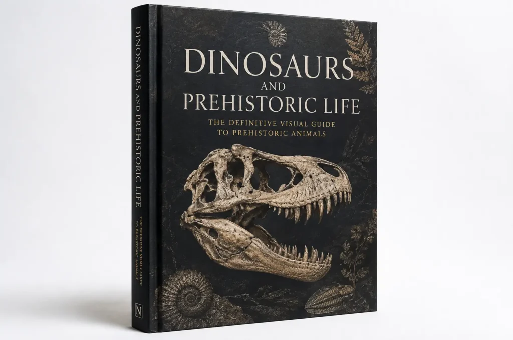
Dinosaurs and Prehistoric Life
A richly illustrated visual reference exploring dinosaurs, fossils, evolution, and prehistoric life across geological time.
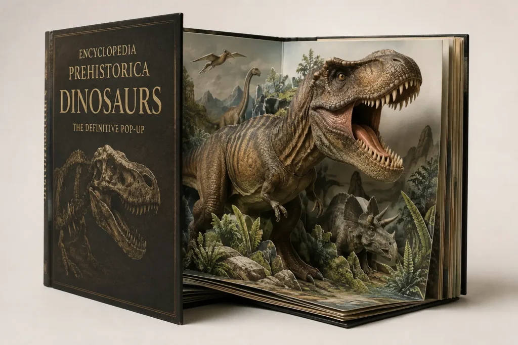
Encyclopedia Prehistorica: Dinosaurs
An interactive dinosaur book featuring spectacular three-dimensional pop-ups, particularly suited to young readers and families.
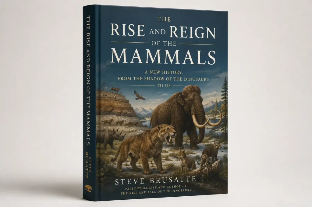
The Rise and Reign of the Mammals
A sweeping history of mammalian evolution, from their origins alongside dinosaurs through the Cenozoic and the emergence of humans.